Abstract

Intercellular adhesion molecule 1 (ICAM-1/CD54) is a transmembrane glycoprotein considered one of the most important adhesion molecules in leukocyte recruitment. It is encoded by the ICAM1 gene and plays a central role in inflammation. Its critical role in various inflammatory diseases such as ulcerative colitis and rheumatoid arthritis has been well established. Given that neuroinflammation, characterized by microglial activation, is a key factor in neurodegenerative diseases like Parkinson's disease (PD), we investigated whether ICAM-1 plays a role in this progressive neurological disorder and, if so, to elucidate its underlying mechanisms. We specifically focused on the possible interactions between ICAM-1, glial cells, and ferroptosis (an iron-dependent form of cell death recently implicated in PD). We conclude that ICAM-1 has both direct and indirect (via glial cells and T cells) effects on ferroptosis, and further elucidation of these interactions may provide new intervention strategies for this devastating disease.

Keywords: ICAM-1; Parkinson's disease; Ferroptosis; Glial cells; T cells; Neuroinflammation

1. Introduction

Intercellular adhesion molecule 1 (ICAM-1/CD54) is a transmembrane glycoprotein discovered in the 1980s, identified as a ligand for the β2 integrin lymphocyte function-associated antigen 1 (LFA-1, CD11a/CD18), serving as a crucial switch for initiating key adhesion pathways [1,2]. Since then, its critical role in inflammatory responses and various inflammatory diseases has been confirmed [3–9].

Although neurodegenerative diseases, particularly Parkinson's disease (PD), are triggered and/or exacerbated by neuroinflammatory mediators, the association between ICAM-1 and PD has not been fully investigated. The rationale supporting this study includes: first, the involvement of neuroinflammation in PD is well-established [10–13]; second, the comorbidity of depression in PD is widely documented [14–16], and the role of ICAM-1 in late-life depression has been demonstrated [17]; furthermore, ICAM-1 expression has been found in reactive astrocytes from PD patients and MPTP-treated monkeys (a non-human primate PD model) [18]. Therefore, this review aims to provide an exposition of the mechanistic significance of ICAM-1 in PD, particularly in relation to glia-mediated neuroinflammation characterized by ferroptosis (a recently implicated pathway in PD pathology) and T-cell reactivity. Thus, after briefly describing ICAM-1, PD, glial cells, T cells, and ferroptosis, we attempt to present compelling evidence for causal relationships among them, with the hope of discovering new therapeutic targets for PD.

1.1 ICAM-1

The known functions of ICAM-1 involve leukocyte extravasation, and it is described as one of the most important adhesion molecules in leukocyte recruitment [19–21]. Specifically, the ICAM1 gene, located on chromosome 19, is induced in endothelial cells by various cytokines and inflammatory mediators, including tumor necrosis factor-α (TNF-α), nuclear factor-κB (NF-κB), interferon-γ (IFN-γ), interleukin-1β (IL-1β), IL-6, as well as hydrogen peroxide (H₂O₂) and NADPH oxidase (NOX) activity [1,22–27]. ICAM-1 is expressed on the plasma membrane and binds to the β2 integrins LFA-1 and macrophage antigen-1 (MAC-1, CD11b/CD18) expressed on leukocytes [28–30]. ICAM-1–LFA-1/MAC-1 binding mediates leukocyte rolling, crawling, adhesion, and the process of blood cells crossing the intact capillary wall (diapedesis), often accompanied by inflammation [30]. Upon binding to LFA-1/MAC-1, ICAM-1 induces the dissociation of junctional proteins between adjacent endothelial cells, cytoskeletal rearrangement, and endothelial nitric oxide synthase (eNOS) activity, thereby promoting leukocyte transendothelial migration [9,30,31,32]. However, as discussed below, the significance of ICAM-1 extends far beyond leukocyte migration.

ICAM-1 is expressed on neurons, immune cells, endothelial cells, and epithelial cells, among others, but at low levels under basal conditions [33–35]. ICAM-1 has several known ligands, including fibrinogen, mucin 1 (MUC1), CD43, hyaluronic acid, rhinovirus, and Plasmodium falciparum [36–41]. Furthermore, ICAM-1 is involved in various physiological processes, such as T-cell regulation (discussed later), macrophage polarization, cell migration, reactive oxygen species (ROS) production, cancer development, and metastasis [7,26,42–44]. Aldosterone and angiotensin II induce atherosclerosis and hypertension, respectively, through ICAM-1-dependent mechanisms in experimental models [45–47]. Angiotensin II infusion also increases ICAM-1 in human subjects [45]. The role of ICAM-1 in atherosclerosis and cardiovascular diseases has been extensively evaluated and confirmed [8,46–48]. This adhesion molecule plays a fundamental role in intestinal and blood-brain barrier (BBB) permeability, as well as in neuroinflammation [49–53]. Beyond inflammatory repair, ICAM-1 also functions in wound healing and efferocytosis (the clearance of apoptotic cells) [7,54,55].

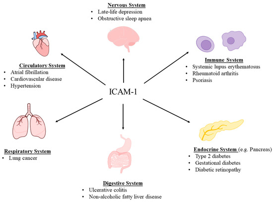
As a transmembrane glycoprotein, ICAM-1 is expressed on the plasma membrane and extends into the cytoplasm and cell surface, enabling it to participate in signal transduction, interact with cytoskeletal structures, and bind ligands [7,30,56]. However, ICAM-1 can be enzymatically cleaved from the cell surface, circulating freely as soluble ICAM-1 (sICAM-1) [57,58]. Specifically, a disintegrin and metalloproteinase 10 (ADAM10), ADAM17, matrix metalloproteinase-2 (MMP-2), and MMP-9 can cleave membrane-bound ICAM-1 [59–62]. Leukocyte elastase and cathepsin G may be particularly relevant in cleaving ICAM-1 isoforms produced by alternative splicing [63,64]. Additionally, sICAM-1 can also be generated through alternative splicing, where it lacks the transmembrane and cytoplasmic domains [30,65]. In vitro studies have found that sICAM-1 concentrations are quantitatively correlated with cell surface ICAM-1 expression [58,66]. Conversely, alternative splicing, protease activity, and shedding of the ICAM-1 extracellular domain may affect this relationship and should be considered in in vivo analyses [30,65,67]. Serum sICAM-1 concentrations in the general population are reported to range from 100 to 450 ng/mL [4,68]. Elevated sICAM-1 levels are associated with various diseases, particularly endometriosis, systemic lupus erythematosus, rheumatoid arthritis, psoriasis, obstructive sleep apnea, non-alcoholic fatty liver disease, lung cancer, atrial fibrillation, obesity, type 2 diabetes, diabetic retinopathy, gestational diabetes, and late-life depression [17,69–82]. Recently, its critical role in ulcerative colitis, an inflammatory bowel disease, was revealed, with higher ICAM-1 levels correlating with poorer prognosis [83] (Figure 1).

Figure 1. ICAM-1 has been implicated in and associated with a variety of diseases.

1.2 Parkinson's Disease (PD)

PD is a progressive neurodegenerative disease characterized by the gradual degeneration of dopaminergic (DAergic) neurons in the substantia nigra pars compacta (SNpc), the accumulation of misfolded α-synuclein into Lewy bodies, and glial cell dysregulation (discussed below). Although both genetic and environmental factors are known risk factors, the majority of PD cases are sporadic or idiopathic (of unknown cause). PD is characterized by motor and non-motor symptoms. Motor symptoms include resting tremor, bradykinesia, rigidity or stiffness, dystonia, and postural and gait abnormalities [11,84]. Freezing of gait is also common. Non-motor symptoms often precede motor symptoms and include partial or complete loss of smell (anosmia), mood disorders (e.g., depression), excessive sweating, hypotension, fatigue, cognitive impairment, inability to make facial expressions or recognize others' verbal and non-verbal cues, sleep disturbances (e.g., insomnia/hypersomnia), gastrointestinal issues (e.g., dysphagia, constipation, and nausea), and urinary and sexual dysfunction [11,85].

Since dopamine (DA) deficiency is the primary underlying cause, therapeutic interventions focus on replenishing this neurotransmitter or its function. This is primarily achieved by administering levodopa (L-dopa, the gold standard) along with drugs that interfere with DA breakdown, such as monoamine oxidase inhibitors or catechol-O-methyltransferase inhibitors (e.g., selegiline/rasagiline and tolcapone/entacapone) and/or newer non-ergot DA agonists (e.g., pramipexole, ropinirole, rotigotine, and apomorphine) [10,86]. Carbidopa is combined with levodopa to prevent its peripheral breakdown. All these drugs significantly alleviate symptoms but none halt the neurodegenerative progression. Furthermore, the most effective, levodopa, not only loses efficacy after months or years of use but can also induce severe dyskinesias, sometimes worse than the initial tremor [10,87]. Therefore, there is an urgent need for more effective interventions without such adverse effects [11,88].

Neuroinflammation and oxidative stress are widely implicated in PD [11,89–92]. Oxidative stress (OS) arises from an imbalance between oxidants and antioxidant capacity, leading to oxidant-induced damage to DNA, proteins, and lipids [11]. In the context of PD, oxidants may originate from various sources, including but not limited to mitochondrial dysfunction, DA metabolism, and glial cells [11,89–92]. Oxidative stress and ROS can induce immune cell activation and immune responses, ultimately leading to inflammation. However, inflammation and OS have a bidirectional relationship, potentially creating a vicious cycle in PD [11,89–92]. Mitochondrial damage and NOX also play roles in this process [90–92]. The focus of this review is to elucidate how ICAM-1 interacts with intermediate mediators of OS or inflammation and how it might serve as a therapeutic target for PD.

1.3 Glial Cells

Glial cells were first discovered in the mid-19th century and were termed neuroglia because they were initially thought to provide only structural support for neurons. However, it is now known that glial cells perform a multitude of critical functions, not only providing structural support for neurons [93–95] but also being involved in myelination [96,97], energy and metabolic control [95,98,99], blood-brain barrier formation [100,101], synaptic development and remodeling [102,103], fluid/electrolyte homeostasis control [104], neurotransmitter regulation [105,106], neuroendocrine function [107], detoxification [108,109], and innate immune responses [110,111]. Therefore, it is not surprising that their disturbance or dysregulation can lead to neuropsychiatric and neurodegenerative diseases [13,97,112–116]. Similarly, they may serve as novel targets for neurological disorders [117]. Indeed, it has been suggested that manipulating nicotinic acetylcholine receptors (nAChRs) in these cells could be a viable intervention target for PD [13], mood disorders, and even drug addiction [118].

The four main glial cell types include microglia, astrocytes, oligodendrocytes, and synantocytes or NG2 cells. In the following, after briefly describing each type, we focus specifically on their interactions with ICAM-1 in neuroinflammatory responses.

1.3.1 Microglia

Microglia constitute 10–15% of the cells in the central nervous system (CNS), covering a large volume of the adult brain parenchyma. These cells continuously monitor the environment through the rapid movement of their filamentous pseudopodia and swiftly respond to any injury. They are homologous to peripheral macrophages but are considered the resident immune cells of the CNS [119,120]. They play a crucial role in maintaining brain homeostasis by regulating neurogenesis, the formation and elimination of neuronal synapses, mediating T-cell infiltration into the brain, and most importantly, clearing pathogens and cellular debris [121]. On the other hand, if overactivated, microglia can induce neuroinflammation, leading to neuronal damage or death, as well as neuropsychiatric and/or neurodegenerative diseases (including PD) [122–125]. A major factor in microglial overactivation is the release of pro-inflammatory mediators (such as IL-1β and IL-6) caused by persistent stress [126–128].

Notably, different microglial subtypes have been previously described based on their activation states. M1 microglia are associated with a pro-inflammatory state, while M2 are associated with an anti-inflammatory state [121,129]. However, new evidence suggests that the functional diversity of microglia stems from their intrinsic properties, and subtypes should be classified based on function, avoiding dichotomies such as M1/M2 [117,130,131].

Microglia express a variety of receptors, including the calcium-sensing receptor (CASR), low-density lipoprotein receptor-related protein 1 (LRP1), triggering receptor expressed on myeloid cells 2 (TREM2), nAChRs, and Toll-like receptors (such as TLR2 and TLR4) [13]. TLRs are a well-characterized family of pattern recognition receptors (PRRs) that initiate innate immune responses by sensing endogenous debris or pathogens. Due to their significant role in neurodegenerative diseases, TLRs are being intensively studied as potential therapeutic targets [121,132–134].

1.3.2 Astrocytes

Named for their star-like morphology, these cells are called astrocytes (astroglia/astrocytes) [135]. They may constitute between 17% and 61% of total brain cells, varying by brain region. Astrocytes also play a key role in maintaining neuronal integrity and function. They provide nutrients, monitor and regulate pH homeostasis, clear waste, and are a critical component of the blood-brain barrier [135,136].

Astrocytes contain glial cell line-derived neurotrophic factor (GDNF), which provides trophic support to neurons, including DAergic neurons [137], and glial fibrillary acidic protein (GFAP), a key protein for maintaining astrocyte strength and the blood-brain barrier. GFAP is often used as a marker for astrocyte identification [138] and may serve as a biomarker for brain and spinal cord diseases [139–142]. These glial cells also express brain-derived neurotrophic factor (BDNF) and the highest amounts of taurine, a free amino acid with antioxidant and anti-inflammatory properties essential for optimal postnatal brain development [143]. It was recently reported that astrocytes are a source of TNF-α, which is necessary for mediating homeostatic synaptic plasticity [144].

Astrocytes and microglia together provide the first line of defense against injury. Here, excessive stimulation of pro-inflammatory signals may synergistically lead to neuronal dysregulation and subsequent neuropsychiatric/neurodegenerative diseases [145–147]. Furthermore, elucidating the close interactions between astrocytes and microglia, as well as between astrocytes and neurons (referred to as crosstalk), may offer new intervention strategies for these diseases [137,148–150].

1.3.3 Oligodendrocytes

Oligodendrocytes (OLs) constitute 75% of all glial cells and are the primary source of CNS myelination [151]. In addition to axonal myelination, OLs have other crucial functions, such as providing metabolic and trophic support through the secretion of GDNF and BDNF, controlling extracellular potassium concentration, and regulating axonal growth [151,152]. They also express TLRs, which are important in myelination [96,153,154]. Therefore, it is not surprising that dysregulation of these glial cells can lead to various neurological disorders, including PD (detailed below).

1.3.4 Syncytial Cells (NG2 Cells)

The fourth major class of glial cells in the CNS are syncytial cells, also known as neuron/glia antigen 2 (NG2) cells and oligodendrocyte precursor cells (OPCs). NG2 cells are expressed in both white and gray matter regions and continue to proliferate in the adult brain [151,155,156]. In addition to being OL progenitor cells, NG2 cells can also transform into astrocytes and neurons [151,155,156]. They have been implicated in various neurological disorders, including multiple sclerosis, Alzheimer's disease (AD), epilepsy, traumatic brain injury, acute ischemic stroke, neurovascular unit formation during development, glioma, and experimental autoimmune encephalomyelitis (EAE) (a disease associated with increased BBB permeability and neuroinflammation) [157–161]. Furthermore, their communication with and influence on neurons make them potential therapeutic targets for various diseases, including PD [162,163], as detailed below.

1.4 Interactions Between ICAM-1 and Glial Cells

1.4.1 ICAM-1 and Microglia

Microglia express ICAM-1 and constitutively express LFA-1 and Mac-1, enabling direct interactions between ICAM-1 and microglia in various contexts [164,165]. Notably, ICAM-1 plays a role in microglial activation [166–168]. Activated microglia, in turn, secrete TNF-α, which induces ICAM-1 expression in vascular endothelial cells and promotes leukocyte infiltration [23,169,170]. ICAM-1 may also indirectly activate microglia. This is because ICAM-1 expression on vascular endothelium facilitates leukocyte transendothelial migration and their infiltration into the CNS, leading to microglial activation [171–174]. Interestingly, leukocytes infiltrating the CNS can adhere to microglia [171,175]. Therefore, a positive feedback loop exists between ICAM-1 and microglia [166–168,171–174].

1.4.2 ICAM-1 and Astrocytes

Astrocytes also contain ICAM-1, and its expression is upregulated by TNF-α, IL-1β, and IFN-γ [176–180]. In turn, ICAM-1 can cause astrocytes to release inflammatory cytokines, including TNF-α [181,182]. ICAM-1 activation of astrocytes can also be achieved indirectly through fibrinogen, which is induced in various neuroinflammatory states and binds to ICAM-1 [183,184]. Fibrinogen-activated astrocytes further enhance ICAM-1 expression and promote NO and ROS production, leading to neuronal death [183,184]. Interestingly, ROS can induce ICAM-1 production in astrocytes via an NF-κB-dependent mechanism [185,186]. Thus, a positive feedback loop also exists here between astrocytes and ICAM-1 [179,181,183–186].

1.4.3 ICAM-1 and Oligodendrocytes

Under inflammatory conditions, enhanced ICAM-1 expression in OLs is considered a defense mechanism against immunogenic injury [177,187]. Direct contact between OLs and T cells is believed to induce OL damage, and anti-ICAM-1 antibodies can inhibit Th1 cell contact with OLs [187]. Due to the role of OLs in myelination, T cell-induced damage to these cells may promote neurodegeneration (as seen in EAE) [187]. The in vivo inhibitory effect of anti-ICAM-1 antibodies on EAE has been demonstrated in animal models, including marmosets [188,189]. As described for microglia, ICAM-1 plays a role in T cell infiltration into the CNS, again providing an indirect mechanism by which ICAM-1 affects OL homeostasis [173,177,187].

1.4.4 ICAM-1 and NG2 Cells

Pro-inflammatory cytokines inhibit the maturation of NG2 cells [190–192]. Furthermore, microglia can influence the proliferation, differentiation, migration, and apoptosis of NG2 cells, while NG2 cells can regulate microglial homeostasis and activation [193]. This suggests an indirect interaction between ICAM-1 and NG2 cells. Although recent findings indicate that NG2 cells participate in the initiation of neuroinflammation by activating immunogenic cells, the NG2 protein appears to be a negative regulator of ICAM-1 expression in pericytes and two different glioblastoma cell lines [194–196].

1.5 ICAM-1—Glial Cells—PD

Neuroinflammation has been widely implicated in PD pathophysiology, with glial cells making significant contributions [197,198]. A recent meta-analysis supports the role of glial cells in neuroinflammation and PD, reporting elevated concentrations of TNF-α, IL-6, IL-1β, nitric oxide (NO), chemokine ligand 2 (CCL2), and C-reactive protein (CRP) in the cerebrospinal fluid (CSF) of PD patients [199]. While transiently activated microglia can be protective, the chronic activation of microglia is widely believed to be involved in PD [200]. Specifically, an inflammatory microglial phenotype has been identified in experimental models and in the substantia nigra (SN) of PD patients [200,201]. Cytokines associated with the inflammatory microglial phenotype include TNF-α, IL-6, IL-1β, and IFN-γ, all of which can induce ICAM-1 expression [1,22,23,199,202]. The role of microglia in PD is further supported by postmortem analyses, which found increased activated microglia and elevated expression of ICAM-1, LFA-1, TNF-α, and IL-6 in the SN and multiple brain regions of PD patients [203].

While microglia have a well-defined role in PD-associated neuroinflammation, they can also induce a neurotoxic and reactive astrocyte phenotype (often termed the A1 phenotype), which may exacerbate PD pathology [204–206]. Furthermore, astrocytes play a role in clearing dysfunctional proteins, such as α-synuclein. Microglia-astrocyte interactions may also facilitate α-synuclein clearance, and its accumulation can increase the expression of ICAM-1 and IL-6 in astrocytes [206–209]. Additionally, increased concentrations of astrocytes and microglia, leukocyte infiltration, and elevated expression of ICAM-1 and LFA-1 have been found in the SN of PD patients [18].

Myelin abnormalities and reduced content are associated with PD symptoms [210,211]. In PD patients, 80% of connections from the basal ganglia show reduced myelin content [212]. Furthermore, a reduction in OLs (the primary contributors to myelination) has been found in idiopathic PD [213]. Additionally, the transfer of α-synuclein from neurons to OLs may exacerbate PD pathology [214,215]. Given that OLs exhibit enhanced ICAM-1 expression under inflammatory conditions, modulating this adhesion molecule may offer a novel target for PD [177,187].

In summary, the above provides a strong link between ICAM-1, glial cells, and PD.

1.6 ICAM-1 and T Cells

Leukocytes in the circulatory system are recruited to sites of inflammation via various inflammatory signaling molecules, such as cytokines and chemokines. As mentioned earlier, upon reaching the inflammatory site, leukocytes often undergo extravasation and diapedesis, i.e., crossing the capillary wall, a process mediated by adhesion molecules. T cells undergo a similar process, as they express LFA-1 and interact with ICAM-1 to facilitate their transendothelial migration [216,217]. ICAM-1 not only promotes T cell transendothelial migration [216,218] but also their activation [219,220]. Furthermore, it plays a crucial role in facilitating interactions between T cells and other leukocytes [219–221].

1.7 ICAM-1—T Cells—PD

T cells play various roles in PD and appear to be influenced by DA [222]. Furthermore, peripheral T cell subset concentrations are typically highly heterogeneous and depend on multiple patient characteristics, such as sex, age, disease severity, and disease duration [223,224]. Specifically, PD patients exhibit increased Th1 and Th17 cells, decreased Th2 and regulatory T cells (Tregs) [224], and some PD patients have α-synuclein-specific T cells [225–227].

Under inflammatory conditions, CNS endothelial cells express various proteins and adhesion molecules (including ICAM-1), promoting the migration and infiltration of immune cells and antibodies [225,228]. T cell infiltration in the CNS of PD patients is supported by numerous animal studies (including non-human primates) and postmortem human studies [229–234]. For example, studies have shown increased expression of ICAM-1 and LFA-1 in endothelial cells and T cells, respectively, and administration of ICAM-1 or LFA-1 antibodies can reduce immunological and behavioral changes in MPTP-treated mice [231]. Furthermore, contact between CD8⁺ T cells and dopaminergic cells has been observed in postmortem examinations of PD patients [231,233]. The ICAM-1/LFA-1 axis has also been shown to mediate Th17-induced dopaminergic neuronal death [35]. Therefore, it can be asserted that the interaction between ICAM-1 and T cells is part of PD pathology. As mentioned earlier, increased ICAM-1 expression in PD is likely most relevant in endothelial cells and the blood-brain barrier, as well as in glial cells [18,203,235–238]. Regarding circulating sICAM-1 concentrations, although ICAM1 decreased gene expression has been detected [239], elevated sICAM-1 levels have also been observed in the serum, plasma, and CSF of these patients [231,240–242]. Therefore,ICAM1 how gene expression translates into protein production in PD remains to be determined.

2. Iron and Ferroptosis

Ferroptosis is an iron-dependent form of regulated cell death, distinct from other cell death mechanisms [243,244]. Although the term and concept of 'ferroptosis' were proposed in 2012, the central role of iron in non-apoptotic cell death emerged as early as 2008 [244,245]. Since then, ferroptosis has been implicated in various diseases, including liver, kidney, intestinal, lung, heart, blood cell, and nervous system disorders [246–248]. Iron (Fe²⁺) reacts with hydrogen peroxide (H₂O₂) to generate hydroxyl radicals (•OH), inducing lipid peroxidation, known as the Fenton reaction (Fe²⁺ + H₂O₂ → Fe³⁺ + •OH + OH⁻) [244]. This reaction and the hydroxyl radicals ultimately lead to lipid peroxidation of cell membranes and cell death (ferroptosis) [244,246,249]. Iron can also react with lipid hydroperoxides to generate alkoxy radicals and activate arachidonic acid lipoxygenase (ALOX) enzymes [246,249]. ALOX enzymes oxidize polyunsaturated fatty acids (PUFAs), generating lipid hydroperoxides, which in turn produce malondialdehyde (MDA) and 4-hydroxynonenal. Due to the role of PUFAs in ferroptosis, PUFA synthases ACSL4 and LPCAT3 are also implicated in this process [249].

H₂O₂ can originate from various pathways, including the reduction of superoxide (O₂⁻) by superoxide dismutase (SOD) [250]. DA metabolism by monoamine oxidase B also produces H₂O₂ [251]. Mitochondria are a major source of superoxide, as electrons can escape from the electron transport chain and react with oxygen [250]. NOX enzymes, which utilize NADPH as an electron donor to produce superoxide, are also a significant source of this radical [249,250]. After superoxide is reduced by SOD, catalase can catalyze the reduction of two H₂O₂ molecules to water and diatomic oxygen [250]. However, as mentioned earlier, in the presence of iron, hydrogen peroxide can participate in the Fenton reaction and contribute to the synthesis of hydroxyl radicals, which are extremely potent oxidants [244].

Ferroptotic cells exhibit several key characteristics, including mitochondrial abnormalities. While iron plays a central role, lipid metabolism and glutathione homeostasis are also regulators of ferroptosis. Similarly, glutathione peroxidase 4 (GPX4) is a primary regulator of ferroptosis [244]. GPX4 utilizes glutathione (GSH) to reduce peroxidized phospholipids and cholesterol [246]. Due to the importance of glutathione, the cystine-glutamate antiporter xCT (also known as SLC7A11) plays a role in mediating ferroptosis. Furthermore, SLC7A11 transports cystine into the cytoplasm while exporting glutamate to the extracellular space. In an NADPH-dependent mechanism, cystine is subsequently converted to cysteine, the rate-limiting amino acid for glutathione synthesis. Notably, the small molecule compound erastin can induce ferroptosis by inhibiting SLC7A11 [252]. The expression of GPX4 and SLC7A11 is regulated by the transcription factor nuclear factor erythroid 2-related factor 2 (Nrf2), which is sequestered and regulated by KEAP1 and binds to antioxidant response elements (AREs) [252,253]. Therefore, Nrf2 is a significant mediator of ferroptosis and regulates the expression of other ferroptosis-related genes, such as glutathione synthetase, ferroportin 1 (FPN1), heme oxygenase 1 (HO-1), transferrin receptor (TFRC), and ferritin heavy chain 1 (FTH1) [253]. Conversely, the transcription factor Bach1 (BTB domain and CNC homolog 1) suppresses the expression of multiple Nrf2-regulated genes, thus potentially inducing ferroptosis by reducing the expression of genes related to glutathione and iron metabolism [252–255]. A specific form of autophagy (known as ferritinophagy) has also recently been revealed to play a significant role in ferroptosis, as it regulates the degradation of ferritin, an intracellular protein that stores and releases iron in a controlled manner. Consequently, ferritinophagy can be viewed as a new player in maintaining iron homeostasis [250,253].

2.1 Ferroptosis and PD

The role of ferroptosis in the pathophysiology of PD has been extensively discussed and is supported by substantial data [85]. Iron can accumulate in the substantia nigra of PD patients, leading to dopaminergic neuronal death [85,256]. PD is also associated with lipid peroxidation, abnormal iron metabolism, reduced GSH, and ROS production, all of which are reflected in differential gene expression in the substantia nigra of PD patients [85,251,257–259]. Specifically, differential expression of ferroptosis-related genes has been observed in dopaminergic and non-dopaminergic neurons, microglia, astrocytes, OLs, NG2 cells, and endothelial cells/pericytes in PD patients [259].

Interestingly, α-synuclein also plays a role in iron metabolism and PUFA synthesis, as it induces lipid peroxidation and increases the risk of ferroptosis in dopaminergic neurons [85,246,260–262]. Iron may also enhance the oxidation of DA (an unstable neurotransmitter), leading to the formation of 6-hydroxydopamine (6-OHDA) and DA quinone (DAQ) [263,264]. DAQ, in turn, may increase neuronal susceptibility to ferroptosis by promoting GPX4 degradation [264,265]. It has also been suggested that Fe³⁺ may be reduced by lipid hydroperoxides, forming an iron-DA complex that produces 6-OHDA and hydroxyl radicals [263,266]. 6-OHDA has been found to increase free iron concentration by releasing iron from ferritin, potentially ultimately creating a vicious cycle of radical generation. This toxic consequence is further enhanced by H₂O₂ production resulting from 6-OHDA metabolism [263].

Ferroptosis may also play a role in blood-brain barrier (BBB) disruption and dysfunction [85,267]. Specifically, increased iron, lipid peroxidation, and decreased antioxidant concentrations have been found in the BBB of PD patients [267]. BBB damage (which may also involve α-synuclein) has been observed in PD patients [268–271] and involves disruption of tight junction proteins and adhesion molecules, thereby promoting disease pathology [267,270].

2.2 Glial Cells and Ferroptosis

Glial cells have complex and multifaceted interactions with iron metabolism and ferroptosis. They can directly serve as a source of iron in the CNS, as they contain ferritin, whose concentration increases under aging and pathological conditions [263]. Glial cells can also indirectly promote iron influx and inhibit its efflux across the BBB by secreting ceruloplasmin and hepcidin, respectively [263,272]. Hepcidin is a peptide hormone produced in the liver that plays a key role in iron homeostasis [85]. Glial cells may also promote iron accumulation in the CNS through cytokine-induced regulation of ferroportin [273].

Since glial cells can regulate CNS iron homeostasis and are involved in inducing ferroptosis, they may play a crucial role in neurodegenerative processes [274]. Specifically, activated astrocytes may induce neuronal ferroptosis by secreting the CXCL3R ligand CXCL10 and reducing SLC7A11 expression [275]. On the other hand, through BDNF- and Nrf2-dependent mechanisms, astrocytes can protect dopaminergic neurons from ferroptosis [276,277]. A similar situation exists for microglia, where lipopolysaccharide (LPS)-activated microglia may protect neurons from glutamate-induced ferroptosis [278]. The complexity of these interactions is further highlighted by the finding that glial cells themselves can also undergo ferroptosis, thereby promoting neurodegeneration [279]. While NG2 cells are particularly susceptible to ferroptosis, oligodendrocytes (OLs), the cells with the highest iron concentration in the CNS, can protect themselves from ferroptosis by secreting ferritin heavy chain [246,277,280,281].

Furthermore, ferroptosis can activate glial cells through the release of damage-associated molecular patterns (DAMPs) [253,282–284]. DAMPs are molecules released from damaged or dead cells and are considered part of the innate immune response [253,282]. Glial cells also express pattern recognition receptors (PRRs, such as TLRs) that can recognize and activate DAMPs, thereby promoting neurodegeneration [283,284]. However, it is noteworthy that DAMP-mediated microglial activation may have neuroprotective effects in certain contexts [284]. This aligns with the concept that acute activation of microglia can have neuroprotective properties, while chronic activation can lead to neurodegeneration [200,201].

2.3 T Cells and Ferroptosis

In tumor cells, T cells can induce ferroptosis through IFN-γ-mediated inhibition of SLC7A11 and activation of ACSL4 [285–288]. This has become an important innate anti-tumor immune response [285,289]. T cells also have the potential to promote neuronal ferroptosis by increasing the expression of transferrin receptor 1 (TfR1) in neurons [290]. Additionally, the interaction between T cells and ferroptosis is reciprocal, as neuronal ferroptosis can activate T cells [291,292], and T cells themselves can undergo ferroptosis [289]. However, ferroptosis appears to be less immunogenic than other forms of cell death [244,246].

2.4 ICAM-1 and Ferroptosis

To our knowledge, the interaction between ICAM-1 and ferroptosis in different contexts has not been extensively studied. Multiple experimental analyses suggest a direct bidirectional interaction between ferroptosis and ICAM-1. For example, in an in vivo model of contusive spinal cord injury, the ferroptosis inhibitor SRS 16-86 reduced ICAM-1 expression along with changes in other protein and cytokine expressions [293]. Similar results were obtained in a rat model of diabetic neuropathy, where SRS 16-86 lowered ICAM-1 as well as IL-1β and TNF-α [294]. Similarly, the ferroptosis inhibitor ferrostatin-1 (Fer-1) inhibited oxidized low-density lipoprotein-induced ICAM-1 expression in endothelial cells [295–297]. Furthermore, the ferroptosis inducer erastin was found to increase ICAM-1 expression and activate endothelial trans-migration [298]. Nonetheless, further validation of the direct interaction between ICAM-1 and ferroptosis in other models and contexts is needed.

Hydrogen peroxide, lipid peroxides, and reactive oxygen species (ROS), which play central roles in ferroptosis, also appear to be involved in ICAM-1 expression [23,26,236,299–302]. Specifically, hydrogen peroxide has been found to increase ICAM-1 expression in endothelial cells [23,299,300]. It should be noted, however, that some analyses failed to detect H₂O₂-induced ICAM-1 expression in endothelial cells [169,303], possibly due to methodological differences [23,169,299,300,303]. Nevertheless, H₂O₂ seems to increase ICAM-1 expression through ICAM1 AP-1 and Ets cis-regulatory elements in the gene promoter [23,299]. H₂O₂ has also been shown to play a role in the post-translational modification of ICAM-1 [304–306]. Additionally, plasma from women with severe preeclampsia, containing high levels of lipid peroxides, increased ICAM-1 expression in human umbilical vein endothelial cells [302].

A recent study indicated a regulatory role for ICAM-1 in ferroptosis, showing that administration of recombinant ICAM-1 (rICAM-1) increased intracellular ROS and Fe²⁺ and decreased GPX4 and SLC7A11 expression in LPS-stimulated macrophages and human umbilical vein endothelial cells, potentially mediated by PTGS2. Furthermore, inhibition of PTGS2 suppressed the effects of rICAM-1 on ferroptosis-related parameters, suggesting a mechanistic role for PTGS2 in this interaction [307]. However, the mechanisms of ICAM-1 in its various roles require further elucidation.

2.5 ICAM-1—Glial Cells—T Cells—Ferroptosis—PD

In PD, DA oxidation and mitochondrial dysfunction are widely considered fundamental features of the disease. DA oxidation appears to play a role in inducing mitochondrial dysfunction, including in sporadic PD cases [308,309]. Mitochondrial dysfunction in both neurons and microglia themselves can induce microglial activation, leading to the release of inflammatory cytokines (such as TNF-α and IL-1β), ultimately causing neuroinflammation and neurodegeneration [310,311]. This self-sustaining cascade of events was proposed two decades ago, although the details of each step were not fully understood at the time [312]. It is now known that inflammatory cytokines released by microglia disrupt the blood-brain barrier and induce the expression of adhesion molecules (such as ICAM-1), thereby promoting the infiltration of leukocytes, including T cells [310]. Once infiltrated, T cell differentiation is influenced by different glial cells (especially microglia) through cytokine release stimulation. Consequently, naïve T cells differentiate into Th1 and Th17 cells, while their differentiation into regulatory T cells is suppressed [310,313]. In turn, CD8⁺ T, Th1, and Th17 cells release inflammatory cytokines, further promoting the transformation of microglia into inflammatory and neurotoxic phenotypes [310]. IL-17 has been found to increase the expression of adhesion molecules in microglia [310,314]. Therefore, this vicious cycle of mutual activation between glial cells and T cells is believed to promote the self-sustaining activation of neuroinflammation and neurodegeneration in PD [198,230,310].

While α-synuclein may promote blood-brain barrier disruption [271,315], T cell infiltration appears to precede α-synuclein accumulation in the brain [233]. Interestingly, elevated ICAM-1 concentration in the CSF of PD patients correlates with increased α-synuclein concentration in the CSF [242]. Once α-synuclein begins to accumulate in the SN, the susceptibility of neurons to ferroptosis increases [85,246,260–262]. This coincides with the presence of activated glial cells and T cells and enhanced ferroptosis [170,260,261,267,273,310]. Neuronal ferroptosis, in turn, activates T cells and glial cells, further propagating the inflammatory and degenerative cycle [283,291,292]. In this context, the abundance of ICAM-1 in the SN may promote T cell-induced dopaminergic neuronal death and further enhance the interaction between glial cells and T cells [18,35,166–168,203].

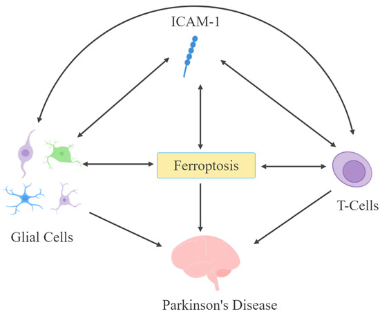
ICAM-1 expression in astrocytes may be enhanced by α-synuclein [207]. Furthermore, ICAM-1-expressing astrocytes are present in the SN and may promote their own ICAM-1 expression through ROS- and NF-κB-dependent mechanisms [18,183–186]. However, to date, inflammatory conditions enhance ICAM-1 expression through cytokine release and can lead to ferroptosis [1,22,23,177,316]. Therefore, a vicious cycle may arise: ferroptosis leads to increased ICAM-1 expression in endothelial cells, causing blood-brain barrier disruption and promoting T cell infiltration, resulting in more cytokine release, neuroinflammation, and neurodegeneration [85,267,293,298,310]. These direct and indirect interactions between ICAM-1, glial cells, T cells, and ferroptosis not only elucidate potential mechanisms leading to PD pathophysiology (Figure 2) but may also offer new intervention strategies (as described below).

Figure 2. ICAM-1, glial cells, T cells, and ferroptosis may influence the pathophysiology of Parkinson's disease through bidirectional interactions.

The association of increased iron accumulation, lipid peroxidation, ROS, elevated ICAM-1, and reduced GSH in the SN of PD patients further supports the existence of a potential direct ICAM-1–ferroptosis axis in the disease [18,85,203,251,257,258,310]. Although indirect, various forms of exercise (recently advocated as potential modulators of ferroptosis) are associated with reduced lipid peroxidation, H₂O₂, iron accumulation, and sICAM-1 concentrations in PD patients [317–320].

3. Novel Intervention Strategies

The urgent need for novel therapeutic approaches for PD is widely recognized [11,88]. In this regard, ICAM-1 and the ICAM-1–ferroptosis axis may represent promising new targets for PD. The internalization of ICAM-1 in endothelial cells following ICAM-1 antibody binding and its subsequent recycling back to the plasma membrane have been documented [321]. ICAM-1 antibodies possess anti-inflammatory potential by inhibiting leukocyte interactions [321] and have been shown in vivo to alleviate PD pathology and symptoms [231,322]. For instance, ICAM-1 antibodies reduce dopaminergic cell death, glial activation, gut dysbiosis, and behavioral alterations in MPTP-treated mice [322]. Similarly, in a previously discussed analysis, LFA-1 and ICAM-1 antibodies reduced immunological and behavioral changes in MPTP-treated mice [231]. Furthermore, inhibition of ICAM-1 or LFA-1 was also found to reduce Treg concentrations in the SN of MPTP-treated mice [323]. Additionally, ICAM-1 antibodies conjugated to catalase have been found in multiple analyses to inhibit H₂O₂ toxicity in endothelial cells [321,324,325]. The ability of antioxidant enzyme-conjugated ICAM-1 antibodies to mitigate various neurological disorders, including glial activation, in experimental traumatic brain injury has been confirmed in vivo [326,327]. Thus, a substantial body of preclinical research confirms the utility of ICAM-1 antibodies in mitigating toxic or neurodegenerative processes.

Furthermore, the F(ab')2 fragment of a murine ICAM-1 antibody was shown to inhibit EAE and, unlike the murine IgG2a ICAM-1 monoclonal antibody, the F(ab')2 fragment did not activate human neutrophils in vitro [188,328]. While the extracellular adherence protein (Eap) of Staphylococcus aureus interacts with multiple ligands, it binds to ICAM-1 and inhibits ICAM-1/LFA-1 interaction, and has been shown to inhibit EAE [329]. Modulating NG2 protein expression may also represent a viable target for regulating ICAM-1 expression [194]. ICAM-1 is also highly expressed in various cancer cells, and ICAM-1 antibodies conjugated to anticancer drugs have recently been evaluated in vivo as a novel approach for cancer therapy [330,331]. Although these novel approaches targeting ICAM-1 have not yet been considered in the context of PD (except for ICAM-1 antibodies), existing data suggest the potential utility of these targets.

The role of levodopa in oxidative stress has been controversial [332–335]. Under physiologically relevant conditions, it appears to have antioxidant activity [333,335]. However, elevated plasma sICAM-1 concentrations were found in idiopathic PD patients (stages 1 and 2) receiving levodopa treatment, suggesting a particular relevance of ICAM-1 early in levodopa therapy [240]. Levodopa-induced dyskinesia was found to coincide with increased inflammatory cytokines and ROS and was exacerbated in the presence of systemic inflammation in vivo [336,337]. Therefore, combining dopamine-enhancing therapy with anti-ICAM-1 therapy could not only target multiple key pathophysiological mechanisms in PD but may also synergize with existing approaches by mitigating side effects. In summary, innovative approaches targeting ICAM-1 and/or the ICAM-1–ferroptosis axis may offer promising options for the treatment and/or mitigation of PD.

4. Conclusion

Recent findings indicate that ICAM-1 plays a central role in PD pathology, manifested through the activation of glial cells and the activation and migration of T cells. Since both glial cells and T cells are directly linked to ferroptosis, this suggests an indirect connection between ICAM-1 and ferroptosis. ICAM-1 may also have direct interactions with ferroptosis, which could occur in the context of PD. Although this latter link requires further confirmation, overall, the existing knowledge supports ICAM-1 as a promising therapeutic target for PD.


Funding: This study was partially supported by NIH/NIGMS (2 SO6 GM08016-39) (YT).

Data Availability Statement: No new data were generated. All citations are available online.

Conflict of Interest: The authors declare no conflict of interest.

Abbreviations (Full Name Translation)

6-OHDA: 6-Hydroxydopamine
AD: Alzheimer's Disease
ADAM10: A Disintegrin And Metalloproteinase 10
ADAM17: A Disintegrin And Metalloproteinase 17
ALOX: Arachidonate Lipoxygenase
ARE: Antioxidant Response Element
BBB: Blood-Brain Barrier
BDNF: Brain-Derived Neurotrophic Factor
CASR: Calcium-Sensing Receptor
CCL2: Chemokine (C-C motif) Ligand 2
CD43: Cluster of Differentiation 43
CNS: Central Nervous System
CRP: C-Reactive Protein
CSF: Cerebrospinal Fluid
DA: Dopamine
DAergic: Dopaminergic
DAMPs: Damage-Associated Molecular Patterns
DAQ: Dopamine Quinone
EAE: Experimental Autoimmune Encephalomyelitis
eNOS: Endothelial Nitric Oxide Synthase
Fer-1: Ferrostatin-1
FPN1: Ferroportin 1
FTH1: Ferritin Heavy Chain 1
GDNF: Glial Cell Line-Derived Neurotrophic Factor
GFAP: Glial Fibrillary Acidic Protein
GPX4: Glutathione Peroxidase 4
GSH: Glutathione
H₂O₂: Hydrogen Peroxide
HO-1: Heme Oxygenase 1
ICAM1: Intercellular Adhesion Molecule 1 (gene)
ICAM-1: Intercellular Adhesion Molecule 1 (protein)
IL-1β: Interleukin-1β
IL-6: Interleukin-6
IFN-γ: Interferon-gamma
LFA-1: Lymphocyte Function-Associated Antigen 1
LPS: Lipopolysaccharide
LRP1: Low-Density Lipoprotein Receptor-Related Protein 1
MAC-1: Macrophage Antigen 1
MDA: Malondialdehyde
MMP-2: Matrix Metalloproteinase-2
MMP-9: Matrix Metalloproteinase-9
MPTP: 1-Methyl-4-phenyl-1,2,3,6-tetrahydropyridine
MUC1: Mucin 1
nAChRs: Nicotinic Acetylcholine Receptors
Nf-kB: Nuclear Factor kappa B
NG2: Neuron-Glial Antigen 2
NO: Nitric Oxide
NOX: NADPH Oxidase
Nrf2: Nuclear Factor Erythroid 2-Related Factor 2
OLs: Oligodendrocytes
OPCs: Oligodendrocyte Precursor Cells
OS: Oxidative Stress
PD: Parkinson's Disease
PRRs: Pattern Recognition Receptors
PUFAs: Polyunsaturated Fatty Acids
ROS: Reactive Oxygen Species
sICAM-1: Soluble Intercellular Adhesion Molecule 1
SN: Substantia Nigra
SNpc: Substantia Nigra Pars Compacta
SOD: Superoxide Dismutase
TfR1: Transferrin Receptor 1
TFRCs: Transferrin Receptors
TLR2: Toll-like Receptor 2
TLR4: Toll-like Receptor 4
TLRs: Toll-like Receptors
TNF-α: Tumor Necrosis Factor Alpha
TREM2: Triggering Receptor Expressed on Myeloid Cells 2
VCAM-1: Vascular Cell Adhesion Molecule 1

Original article available at:Intercellular Adhesion Molecule 1 (ICAM-1): An Inflammatory Regulator Potentially Involved in Ferroptosis and Parkinson's Disease.pdf